Pneumothorax Ultrasound - Pneumothorax on shoulder X-ray - Radiology at St. Vincent's University Hospital - Supine pneumothorax) for the identification of pneumothorax after blunt trauma.. There is normal lung sliding on the right side. Since then there have been many studies that have shown bedside ultrasound can rapidly detect pneumothorax, helping avoid serious potential consequences (i.e. Identification of a lung point on lung us yields 100% specificity for pneumothorax (58). In a pneumothorax, since there is no movement, using m mode will. Pneumothorax can be missed by bedside radiography, and computed tomography is the current alternative.
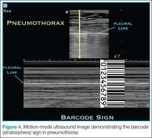
In a pneumothorax, since there is no movement, using m mode will. Pneumothorax can be missed by bedside radiography, and computed tomography is the current alternative. Remember air in pneumothorax will typically rise to the least dependent portion of the hemithorax. Ultrasound proved more sensitive than bedside radiography. Nov 08, 2017 · primary spontaneous pneumothorax:
Emergency Ultrasound: Pneumothorax Assessment | MDedge Emergency Medicine from cdn.mdedge.com Each site was scanned twice at each time point. Identification of a lung point on lung us yields 100% specificity for pneumothorax (58). A pneumothorax can be caused by: Place a linear (vascular/soft tissue) probe in the most anterior point of the chest wall, usually at about the 3rd or 4th intercostal space. The presence of a 'lung point' indicates pneumothorax. These are the thoracic radiographs: Ultrasound scans in all 43 examinable patients with pneumothorax showed absent lung sliding, 41 of 43 patients had the a line sign, and 34 exhibited a lung point. A recent review article by wilcox in jama questions whether ultrasound guidance truly reduces the risk of pneumothorax.
A pneumothorax can develop into a collapsed lung.
A pneumothorax separates the visceral and parietal pleura, eliminating normal lung sliding between these layers on lung us. Once you understand these basic lung ultrasound findings you will be able to interpret just about any lung ultrasound images. When m mode is used on a normal lung, the lung (and the air inside it) moves back and forth across this single ultrasound beam creating a picture that is often compared to a sandy beach. Unless the pneumothorax is loculated or there are adhesions, the gas moves freely within the thoracic cavity. There is normal lung sliding on the right side. Lung ultrasound pathology profiles such as pneumothorax, pneumonia, cardiogenic pulmonary edema, etc will have a different combination and distribution of these pathological lung ultrasound findings/signs. Nov 08, 2017 · primary spontaneous pneumothorax: 3 article feature images from this case Ein pneumothorax (gelber pfeil) mit noch geringer. To evaluate for pneumothorax with ultrasound, have the patient lay supine. In the supine trauma patient, this will typically be the anterior chest wall lateral to the sternum in the second intercostal space. Among 302 analyzable controls, 65 had absent lung sliding, 16 of them showed an a line sign, and none showed a lung point. Each site was scanned twice at each time point.
Ultrasound proved more sensitive than bedside radiography. At its heart, the concept behind using ultrasound to evaluate for air in the space between the visceral and parietal pleura is straightforward. Remember air in pneumothorax will typically rise to the least dependent portion of the hemithorax. Pneumothorax therapie universitatsspital zurich from new.usz.ch pleural effusion, pneumothorax and contusion of the lung (18%) are found in more severe cases. There was no hemothorax on the left side.
Chest XRay in Pneumothorax from fpnotebook.com The diagnosis of pneumothorax using ultrasound is accurate and reliable; Supine pneumothorax) for the identification of pneumothorax after blunt trauma. Since then there have been many studies that have shown bedside ultrasound can rapidly detect pneumothorax, helping avoid serious potential consequences (i.e. These are the thoracic radiographs: 3 article feature images from this case Home ultrasound library a pneumothorax is an abnormal collection of gas in the pleural space, separating the parietal pleura of the chest wall from the visceral pleura of the lung. Ultrasound appearance of pneumothorax explained. Pneumothorax can be missed by bedside radiography, and computed tomography is the current alternative.
Differential diagnosis the thin white line of the visceral pleura in pneumothorax must be distinguished from the black line of a skin fold, which is accentuated by the mach effect ( fig.
Ruling out a pneumothorax is the easy part for me, and it doesn't require much more than a quick visual inspection. Ultrasound can also allow semiquantitative assessment of pneumothorax size by assessing the position of the lung point. Once you understand these basic lung ultrasound findings you will be able to interpret just about any lung ultrasound images. · february 14, 2020 · 1 min read. Nov 08, 2017 · primary spontaneous pneumothorax: Ultrasound use may therefore obviate the need for ct in a majority of cases. Additionally, it can result in timely diagnoses specifically in neonatal pneumothorax. The probe should be oriented perpendicular to the ribs (usually marker dot towards the head). You can also use m mode, or motion mode, which provides an image showing tissue motion along a single ultrasound beam. Unless the pneumothorax is loculated or there are adhesions, the gas moves freely within the thoracic cavity. At its heart, the concept behind using ultrasound to evaluate for air in the space between the visceral and parietal pleura is straightforward. Ultrasound scans in all 43 examinable patients with pneumothorax showed absent lung sliding, 41 of 43 patients had the a line sign, and 34 exhibited a lung point. Ultrasound outperforms cxr in evaluation of pneumothorax in blunt trauma patients, but there cite this article as:
A recent review article by wilcox in jama questions whether ultrasound guidance truly reduces the risk of pneumothorax. Spontaneous pneumothorax in a cat. 3 article feature images from this case There was no hemothorax on the left side. Lung ultrasound pathology profiles such as pneumothorax, pneumonia, cardiogenic pulmonary edema, etc will have a different combination and distribution of these pathological lung ultrasound findings/signs.
SonoTip&Trick: There's a left pneumothorax! Really? check again… | SonoSpot: Topics in Bedside ... from img.youtube.com This is where we want to look with our ultrasound. Once you understand these basic lung ultrasound findings you will be able to interpret just about any lung ultrasound images. Ultrasound use may therefore obviate the need for ct in a majority of cases. 1 traumatic pneumothorax is common in dogs, whereas spontaneous pneumothorax is relatively rare. Supine pneumothorax) for the identification of pneumothorax after blunt trauma. Keine äußere verletzung des brustkorbs. A recent review article by wilcox in jama questions whether ultrasound guidance truly reduces the risk of pneumothorax. A pneumothorax can be caused by:
Remember air in pneumothorax will typically rise to the least dependent portion of the hemithorax.
Nov 08, 2017 · primary spontaneous pneumothorax: Ultrasound outperforms cxr in evaluation of pneumothorax in blunt trauma patients, but there cite this article as: Lack of ionizing radiation and easy operation are benefits of this imaging technique. 1 traumatic pneumothorax is common in dogs, whereas spontaneous pneumothorax is relatively rare. In the supine trauma patient, this will typically be the anterior chest wall lateral to the sternum in the second intercostal space. When m mode is used on a normal lung, the lung (and the air inside it) moves back and forth across this single ultrasound beam creating a picture that is often compared to a sandy beach. To evaluate for pneumothorax with ultrasound, have the patient lay supine. A pneumothorax separates the visceral and parietal pleura, eliminating normal lung sliding between these layers on lung us. Among 302 analyzable controls, 65 had absent lung sliding, 16 of them showed an a line sign, and none showed a lung point. Since then there have been many studies that have shown bedside ultrasound can rapidly detect pneumothorax, helping avoid serious potential consequences (i.e. These are the thoracic radiographs: Thoracic ultrasound has more sensitivity than a supine chest radiograph (see: Identification of a lung point on lung us yields 100% specificity for pneumothorax (58).
There is normal lung sliding on the right side pneumothorax. Tension pneumothorax), especially in patients requiring mechanical ventilation.